Unit 1 · Topic 1.7 · about 30 minutes · 7 parts · 12 scored checks, 3 of them multiple choice, plus 2 from earlier lessons
AP Biology 1.7: Proteins
Unit 1: 8 to 11% of the multiple-choice section
What you'll be able to do
- Describe the structure of proteins, from amino acids and peptide bonds through primary, secondary, tertiary and quaternary structure, and how that structure determines a protein's function.
Key vocabulary
| Term | What it means | The exam trap |
|---|---|---|
| Amino acid | The monomer of a protein: a central carbon bonded to a hydrogen, a carboxyl group, an amine group and an R group. | Every amino acid shares three of those four groups, so a question about how amino acids differ is a question about R groups. |
| Peptide bond | The covalent bond between the carboxyl group of one amino acid and the amine group of the next. | Peptide bonds hold the chain together, so a question about folding is usually about other bonds and interactions. |
| R group | The variable side chain of an amino acid, which is hydrophobic, hydrophilic or ionic. | Two different amino acids can share R group properties, so compare the properties, not just the names. |
| Primary structure | The specific sequence of amino acids in a polypeptide. | Primary structure is the order of amino acids, not a shape, so a choice that describes folding is describing a different level. |
| Secondary structure | Local folding of a polypeptide into alpha-helices and beta-pleated sheets. | Exam choices often swap what holds secondary and tertiary structure, so tie each level to the part of the chain involved. |
| Tertiary structure | The full three-dimensional shape of a polypeptide. | Tertiary structure involves several kinds of interaction, so a choice naming only one of them may be incomplete. |
| Quaternary structure | The arrangement of two or more polypeptides in a protein. | A protein of one chain has no quaternary structure, so count the polypeptides before choosing this level. |
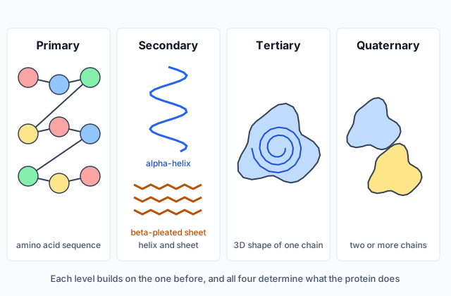 Tap to open the picture full size
Part 1 Think first One amino acid out of place
Commit to a guess first. Being wrong now makes the right idea stick better.
Predict first
In one inherited condition, a person's hemoglobin, the protein that carries oxygen in red blood cells, differs from the usual form at a single amino acid. Every other amino acid in the chain is the same. Yet the person's red blood cells can bend into a stiff sickle shape and block small blood vessels. How can one amino acid matter so much?
In short. A protein is one or more chains of amino acids joined by peptide bonds, and the order of those amino acids, through the interactions of their R groups, folds the protein into the shape that decides its function.
- A peptide bond forms between the carboxyl group of one amino acid and the amine group of the next.
- Each amino acid's R group is hydrophobic, hydrophilic or ionic, and R group interactions shape the protein.
- Primary, secondary, tertiary and quaternary structure all determine what a protein can do.
Part 2 Learn it From a chain of amino acids to a working shape
2a. Amino acids and peptide bonds
Proteins are polymers, and their monomers are amino acids. Every amino acid is built around a central carbon atom with four things covalently bound to it: a hydrogen atom, a carboxyl group (−COOH), an amine group (−NH₂) and a variable R group, or side chain. The first three are the same in every amino acid. The R group is what makes each one different.
Amino acids are joined into a chain by dehydration synthesis (Topic 1.3). A covalent bond, the peptide bond, forms between the carboxyl group of one amino acid and the amine group of the next, and a water molecule is released. Adding amino acid after amino acid this way builds a growing peptide chain, a polypeptide.
Because every amino acid has an amine group, every protein contains nitrogen, and the sulfur from Topic 1.2 is carried in some R groups. The detailed molecular structure of each amino acid is beyond this course.
Quick check · Fill the statement
Complete the description of how two amino acids are joined.
A peptide bond forms between the group of one amino acid and the group of the next. The part of each amino acid that differs from one amino acid to another is its .
2b. R groups decide how a region behaves
R groups fall into three categories by their chemical properties:
- Hydrophobic (nonpolar): they avoid water, like the tails of a lipid.
- Hydrophilic (polar): they attract water and can form hydrogen bonds.
- Ionic: they carry a charge, positive or negative.
The interactions of the R groups determine the structure and function of each region of a protein. In a protein dissolved in a cell's watery interior, hydrophobic R groups tend to cluster together in the middle, away from water, while hydrophilic and ionic R groups tend to sit on the surface, facing the water. Oppositely charged ionic R groups attract each other.
That is how one amino acid can matter. Replace a hydrophobic R group in the protein's water-free core with an ionic one, and that region now has a charge where none fits, so it may fold differently. Replace a charged R group on the surface with a hydrophobic one, and that patch may now stick to hydrophobic patches on other molecules.
Quick check · Sort it
Each card describes an R group in a protein dissolved in water. Tap a card, then the category it most likely belongs to.
Hydrophobic (nonpolar)
Hydrophilic (polar)
Ionic
2c. Primary and secondary structure
Biologists describe a protein's shape in levels.
Primary structure is the specific sequence of amino acids in the polypeptide, the order in which they are joined. That sequence determines the primary structure, and it also determines the overall shape the protein folds into, because it decides which R groups sit where.
Secondary structure is local folding along the chain. It forms from interactions between atoms of the polypeptide backbone, the repeating part of the chain, not the R groups. Hydrogen bonds between backbone atoms coil stretches of the chain into alpha-helices or fold them into beta-pleated sheets.
So a single protein can have helices in one region and sheets in another, each held by backbone hydrogen bonds, all set up by the sequence.
Quick check · Multiple choice
A stretch of a polypeptide coils into an alpha-helix. Which statement describes this structure correctly?
2d. Tertiary and quaternary structure
Tertiary structure is the protein's full three-dimensional shape. It forms from interactions among the R groups: hydrogen bonds, hydrophobic interactions that pull nonpolar R groups together, ionic interactions between charged R groups, and disulfide bridges, covalent bonds between sulfur atoms in two R groups.
Quaternary structure exists only in proteins made of more than one polypeptide. It arises from the interactions between those polypeptides as they fit together. Hemoglobin, for example, is built from several polypeptide chains.
All four levels of structure determine a protein's function. An enzyme's active site, a pocket in its tertiary shape, fits its substrate only if every level is right. A change at the primary level, one amino acid, can travel up through the other levels and change what the protein does.
Quick check · Match
Match each level of protein structure to its description.
Part 3 Watch it done Which change matters?
Predict first, then watch each step, then finish the reasoning yourself.
An enzyme dissolved in a cell's watery interior has a hydrophobic amino acid at position 88, in the core of the folded protein. Two different mutations each change only that amino acid.
- Mutation 1 replaces it with a different hydrophobic amino acid.
- Mutation 2 replaces it with an amino acid whose R group is ionic.
Which mutation is more likely to change the enzyme's shape and activity?
Predict first
Before you open the first step, predict: which mutation is more likely to change the enzyme?
Step 1 · Find where the amino acid sits
Position 88 is in the core of the folded enzyme, away from the water. That is where hydrophobic R groups cluster, held together by hydrophobic interactions that help set the tertiary structure.
Step 2 · Compare the new R groups
Mutation 1 keeps the same category, hydrophobic, so the new R group can join the same hydrophobic interactions. Mutation 2 puts a charged, ionic R group in a place with no water and no partner charge to interact with.
Why this step?
Why does the category of the R group matter more than the exact amino acid?
The interactions that fold a region depend on the R group's chemical properties: hydrophobic, hydrophilic or ionic. An R group with the same properties can make the same kind of interactions, even if it is a different amino acid.
Step 3 · Trace the change up the levels
A changed primary structure changes the R groups in that region. If the new R group cannot make the same interactions, the tertiary structure can shift, and if the active site's shape changes, so does the enzyme's activity.
Your step · Decide and justify
Which mutation is more likely to change the enzyme's shape and activity?
Which reason supports your decision?
One changed amino acid matters when its R group has different properties from the one it replaced, because the interactions that fold a protein, and fit it to other molecules, depend on those properties. Sickle hemoglobin is the surface version: a charged R group on the outside is replaced by a hydrophobic one, and the hemoglobin molecules stick to each other.
Part 4 Try it A protein of four chains
A new protein. A protein is made of 4 polypeptides fitted together. Each polypeptide is a single unbranched chain of 141 amino acids.
Try it · Calculate
How many peptide bonds does the whole protein contain?
Try it · Decide and justify
A student says this protein has quaternary structure. Is the student right?
Which reason supports your decision?
Part 5 Explain it One change, traced through the levels
On the exam this is handwritten, so answer on paper first. Nothing you type here leaves this page. Then check your answer against the model and each scoring point.
Free response is 50% of the exam score. This one is shaped like Question 4, Conceptual Analysis.
Free-response practice · Question 4 style
A protein that works in the watery interior of a cell has a hydrophobic amino acid in its folded core. A mutation replaces it with an amino acid that has an ionic R group. Nothing else in the sequence changes.
(a) Describe the bond that joins the new amino acid to its neighbors in the chain.
(b) Explain how a change in the amino acid sequence can change a protein's overall shape.
(c) Explain how the ionic R group could change the protein's tertiary structure.
(d) Predict the effect of this change on the protein's function, and justify your prediction.
Model answer
(a) A peptide bond, a covalent bond formed between the carboxyl group of one amino acid and the amine group of the next.
(b) The sequence of amino acids determines the primary structure, and the interactions of the R groups along that sequence determine how the chain folds, so a different sequence can fold into a different shape.
(c) The core was held together partly by hydrophobic interactions among nonpolar R groups. A charged R group cannot take part in those interactions, so that region can fold differently and the tertiary structure can change.
(d) The protein's function will likely be reduced or lost, because a protein's function depends on its structure, and a change in tertiary structure can change shapes such as an active site.
Score your own answer. This is a self-check, not a grade. Tick each point your answer makes.
Part 6 Check yourself Changes at one position
All three case questions use the table.
A researcher makes four versions of an enzyme that differ only at position 42, a spot in the enzyme's folded core, and measures each version's activity under the same conditions. These numbers are a teaching example.
| Version | R group at position 42 | Activity (% of normal) |
|---|---|---|
| Normal | Hydrophobic | 100 |
| Version 1 | A different hydrophobic R group | 97 |
| Version 2 | Hydrophilic (polar) | 38 |
| Version 3 | Ionic | 6 |
Case question 1 · Calculate
Using the table: version 2's activity is how many times version 3's? Give your answer to one decimal place.
Case question 2 · Multiple choice
Using the table: why does version 1 keep nearly all of the normal activity?
Case question 3 · Multiple choice
Using the table: a fifth version puts an ionic R group of the opposite charge at position 42. Which prediction is best supported?
The rest stand on their own and do not use the case.
Check yourself · Spot the error
A student describes how a protein folds. Tap the line with the mistake.
Check yourself · Put it in order
Put the steps in building a working protein made of two polypeptides in order.
Part 7 Keep it Bring back earlier ideas
Proteins are polymers like the nucleic acids of Topic 1.6 and the carbohydrates of Topic 1.4. Pulling both back up is what makes them last.
From 1.6 Nucleic Acids · Spot the error
A student compares RNA with DNA. Tap the line with the mistake.
From 1.4 Carbohydrates · Fill the statement
Complete the description of how carbohydrates are built.
The monomers of carbohydrates are , also called simple sugars. They are joined by bonds to form , the complex carbohydrates.
In three lines
- Proteins are chains of amino acids joined by peptide bonds between the carboxyl group of one and the amine group of the next.
- Each R group is hydrophobic, hydrophilic or ionic, and R group interactions, set by the sequence, shape each region of the protein.
- Primary, secondary, tertiary and quaternary structure all determine a protein's function.
Exam strategy for this topic
- Section I, Multiple Choice: 60 questions in 90 minutes.
- Section II, Free Response: 6 questions in 90 minutes.
- This lesson's Explain it is shaped like free-response question 4, Conceptual Analysis.
- A four-function, scientific, or graphing calculator is allowed on both sections of the exam.
When a question changes one amino acid, name the old and new R group categories, say where the change sits, and trace it up the levels: primary, then the folding it changes, then the function.
Questions students ask
How are amino acids joined together?
By peptide bonds, covalent bonds that form between the carboxyl group of one amino acid and the amine group of the next during dehydration synthesis.
What are the three kinds of R group?
Hydrophobic (nonpolar), hydrophilic (polar) and ionic (charged). The interactions of the R groups determine the structure and function of each region of a protein.
What is the difference between secondary and tertiary structure?
Secondary structure is local folding into helices and sheets, held by hydrogen bonds between backbone atoms. Tertiary structure is the whole 3D shape, set by interactions among R groups.
Does every protein have quaternary structure?
No. Quaternary structure arises from interactions between multiple polypeptides, so only proteins made of two or more chains have it.
Why can changing one amino acid change a protein's function?
The sequence determines the protein's shape. A new R group with different properties can change how a region folds, and all four levels of structure determine function.
Beyond the AP Exam
College Board sets this limit on what the AP Exam asks about this topic. Anything past it is not on the exam.
The molecular structure of amino acids is beyond the scope of the AP Exam.
Course alignment, for teachers
AP Biology topic 1.7, Unit 1: Chemistry of Life.
- Skill 6.E: Predict the causes or effects of a change in, or disruption to, one or more components in a biological system.
This lesson maps all 6 essential knowledge statements of the topic to the part that teaches each one: 6 are checked by a scored question. The map is in the lesson file.