Unit 2 · Topic 2.3 · about 30 minutes · 7 parts · 11 scored checks, 2 of them multiple choice, plus 2 from earlier lessons
AP Biology 2.3: Plasma Membrane
Unit 2: 10 to 13% of the multiple-choice section
What you'll be able to do
- Describe the roles of phospholipids and embedded proteins in the cell membrane, and how their hydrophilic and hydrophobic regions are arranged.
- Describe the fluid mosaic model of cell membranes.
Key vocabulary
| Term | What it means | The exam trap |
|---|---|---|
| Phospholipid | A lipid with a polar phosphate region and two nonpolar fatty acid tails; the main molecule of the membrane's framework. | A phospholipid has both kinds of region in one molecule, so name the region you mean when you say whether it is polar. |
| Hydrophilic | Interacting well with water; true of polar and charged regions of a molecule. | One molecule can be hydrophilic in one region and hydrophobic in another, so describe regions rather than whole molecules. |
| Glycoprotein | A protein with carbohydrate chains attached to it. | Glycoproteins and glycolipids both carry carbohydrate, so look at what the carbohydrate is attached to before naming one. |
| Cholesterol | A steroid found in the membranes of vertebrate animals. | Cholesterol is a lipid but not a phospholipid, so do not count it as part of the framework. |
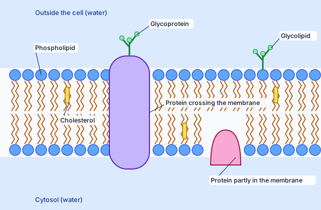 Tap to open the picture full size
Part 1 Think first Two cells become one
Commit to a guess first. Being wrong now makes the right idea stick better.
Predict first
Biologists tag the membrane proteins of a human cell with a red glowing marker and those of a mouse cell with a green one. Then they fuse the two cells, so the new cell starts out with one red half and one green half of membrane. They keep it at body temperature for 40 minutes. What do they most likely see?
In short. The plasma membrane is a framework of phospholipids with proteins, steroids, glycoproteins and glycolipids embedded in it, all able to move, and each part's hydrophilic and hydrophobic regions decide where it sits.
- Phospholipids turn their polar phosphate heads toward the water on each side and their nonpolar fatty acid tails toward each other inside.
- Embedded proteins have hydrophobic surfaces where they meet the tails and hydrophilic regions inside the protein or exposed to the cytosol.
- The fluid mosaic model: many kinds of molecules, all able to move around the surface of the cell within the membrane.
Part 2 Learn it What the membrane is made of
2a. Phospholipids form the framework
The membrane's framework is a double layer of phospholipids. Each phospholipid has two different regions. Its phosphate region is polar and hydrophilic: it interacts well with water. Its two fatty acid tails are nonpolar and hydrophobic: they do not.
A cell has water on both sides of its membrane, the aqueous environment outside and the cytosol inside. So the phospholipids arrange themselves in two rows. The hydrophilic phosphate regions of each row face outward, toward the water on that side. The hydrophobic fatty acid tails of the two rows face each other in the interior of the membrane, where no water reaches them.
That arrangement is what makes the membrane a boundary. Its interior is a thin layer of nonpolar tails between two watery environments.
Quick check · Fill the statement
Complete the description of how phospholipids are arranged in a plasma membrane.
The phosphate regions face the watery environment outside and inside the cell, and the fatty acid tails of the two rows face each other in the of the membrane.
2b. Embedded proteins follow the same rule
Proteins are embedded in the phospholipid framework, and they sit the way they do for the same reason. A protein's amino acids have side groups. Some side groups are charged or polar, so those regions of the protein are hydrophilic. Others are nonpolar, so those regions are hydrophobic. An embedded protein can be hydrophilic, hydrophobic, or both.
The hydrophobic regions make up the protein surface that interacts with the fatty acid tails in the interior of the membrane. A protein that crosses the whole membrane has a band of nonpolar side groups around its middle, right where the tails are.
The hydrophilic regions are either exposed to the cytosol (the fluid part of the cytoplasm) or tucked inside the interior of the protein itself. A protein that lets ions through, for example, can have a hydrophilic passage running down its center, lined with polar side groups, while its outer surface in the membrane stays hydrophobic.
Quick check · Sort it
Each card describes a region of a protein that crosses the plasma membrane. Tap a card, then the kind of region it most likely is.
Hydrophobic region
Hydrophilic region
2c. A fluid mosaic
A plasma membrane holds more than phospholipids and proteins. Its structural framework of phospholipids is embedded with:
- proteins, crossing the membrane or sitting partly in it
- steroids, such as cholesterol in the membranes of vertebrate animals, nestled among the tails
- glycoproteins, proteins with carbohydrate chains attached
- glycolipids, lipids with carbohydrate chains attached
That mix of many kinds of molecules is the "mosaic." The "fluid" part is that all of them can move around the surface of the cell within the membrane. A phospholipid drifts sideways among its neighbors; an embedded protein drifts through the phospholipids. The membrane is held together by how its parts interact with water, not by fixed bonds between them, so it behaves more like a thin liquid than a solid sheet. Together this picture is the fluid mosaic model.
Quick check · Match
Match each membrane component to its description.
Part 3 Watch it done Which stretch crosses the membrane?
Predict first, then watch each step, then finish the reasoning yourself.
A protein crosses the plasma membrane once. Biologists count the amino acids in two stretches of its chain and how many have nonpolar side groups.
- Stretch 1: 20 amino acids, 3 with nonpolar side groups
- Stretch 2: 22 amino acids, 19 with nonpolar side groups
These numbers are a teaching example. Which stretch most likely lies in the membrane's interior?
Predict first
Before you open the first step, predict: which stretch lies in the membrane's interior?
Step 1 · Turn each count into a percentage
Stretch 1: 3 of 20 is 0.15, or 15% nonpolar. Stretch 2: 19 of 22 is 0.864, or about 86% nonpolar.
Why this step?
Why use percentages instead of the counts themselves?
The stretches are different lengths, 20 and 22 amino acids. A percentage compares them on the same scale, so a longer stretch does not look more nonpolar just because it is longer.
Step 2 · Decide what each region must touch
A region in the membrane's interior touches the fatty acid tails, so it should be hydrophobic: mostly nonpolar side groups. A region outside the bilayer touches water, so it should be hydrophilic: mostly charged and polar side groups.
Your step · Decide and justify
Which stretch most likely lies in the membrane's interior?
Which reason supports your decision?
The rule is the same for every part of the membrane: what faces the tails is hydrophobic, and what faces water is hydrophilic. Stretch 2 crosses the bilayer, and stretch 1 most likely hangs in the cytosol or outside the cell.
Part 4 Try it A bleached spot that comes back
A new method, a new question. A biologist attaches a glowing tag to the phospholipids in a cell's plasma membrane. She uses a laser to bleach one small spot, so the tags there stop glowing for good, and then measures the spot's brightness as a percentage of its brightness before bleaching. These numbers are a teaching example.
- Before bleaching: 100%
- Just after bleaching: 10%
- 20 seconds later: 60%
- 60 seconds later: 90%
Try it · Calculate
By 60 seconds, what percentage of the brightness the spot lost to bleaching had it regained? Give your answer to the nearest whole percent.
Try it · Decide and justify
Do these data support the claim that phospholipids move within the membrane?
Which reason supports your decision?
Part 5 Explain it What a membrane drawing leaves out
On the exam this is handwritten, so answer on paper first. Nothing you type here leaves this page. Then check your answer against the model and each scoring point.
Free response is 50% of the exam score. This one is shaped like Question 5, Analyze Model or Visual Representation of a Biological Concept or Process.
Free-response practice · Question 5 style
A textbook drawing of a plasma membrane shows two rows of phospholipids, a protein that crosses both rows, cholesterol among the fatty acid tails, and a carbohydrate chain attached to a protein on the outer surface.
(a) Describe how the phospholipids in the drawing are oriented relative to the water on each side of the membrane.
(b) Explain why the part of the protein that crosses the two rows has mostly nonpolar side groups.
(c) Identify the component formed by a carbohydrate chain attached to a protein.
(d) Explain one way the drawing misrepresents a real membrane, using the fluid mosaic model.
Model answer
(a) The hydrophilic phosphate regions face the water outside the cell and the cytosol inside, and the hydrophobic fatty acid tails of the two rows face each other in the interior.
(b) That part of the protein interacts with the hydrophobic fatty acid tails in the membrane's interior, so its surface must be hydrophobic, which nonpolar side groups make it.
(c) A glycoprotein.
(d) A drawing fixes every molecule in one place, but in a real membrane the phospholipids, proteins, cholesterol, glycoproteins and glycolipids can all move around the surface of the cell within the membrane.
Score your own answer. This is a self-check, not a grade. Tick each point your answer makes.
Part 6 Check yourself Mixing after fusion
All three case questions use the table.
A biologist repeats the experiment from Think first. She fuses human and mouse cells whose membrane proteins carry red and green tags, keeps the fused cells under one of three conditions for 40 minutes, then counts how many show the two colors mixed over the whole surface. In the third condition, a drug stops the cells from making any new proteins. These numbers are a teaching example.
| Condition | Cells counted | Cells with colors mixed |
|---|---|---|
| 37 °C | 200 | 180 |
| 15 °C | 200 | 24 |
| 37 °C, new protein synthesis blocked | 200 | 174 |
Case question 1 · Calculate
Using the table: what percentage of the fused cells kept at 15 °C had mixed colors?
Case question 2 · Multiple choice
Using the table: what does the result with new protein synthesis blocked show?
Case question 3 · Multiple choice
Using the table: which explanation best fits the result at 15 °C?
The rest stand on their own and do not use the case.
Check yourself · Spot the error
A student describes the fluid mosaic model. Tap the line with the mistake.
Check yourself · Decide and justify
A student's drawing of a membrane shows the fatty acid tails of the outer row pointing out into the water around the cell. Is the drawing correct?
Which reason supports your decision?
Part 7 Keep it Bring back earlier ideas
Which R groups are hydrophobic, from Unit 1, decides where a membrane protein sits, and the last lesson's surface area is this membrane's. Pulling them back up is what makes them last.
From 1.7 Proteins · Sort it
Each card describes an R group in a protein dissolved in water. Tap a card, then the category it most likely belongs to.
Hydrophobic (nonpolar)
Hydrophilic (polar)
Ionic
From 2.2 Cell Size · Fill the statement
Complete the comparison of two cells with the same volume.
Cell 1 is round. Cell 2 is long and thin, with folds in its membrane. Cell 2 has the surface area-to-volume ratio, so it can exchange materials efficiently with its environment.
In three lines
- Phospholipids face their hydrophilic phosphate regions toward the water on each side and their hydrophobic fatty acid tails toward each other inside.
- Embedded proteins are hydrophobic where they meet the tails and hydrophilic where they are exposed to the cytosol or inside the protein.
- The fluid mosaic model: proteins, steroids, glycoproteins and glycolipids in a phospholipid framework, all able to move within the membrane.
Exam strategy for this topic
- Section I, Multiple Choice: 60 questions in 90 minutes.
- Section II, Free Response: 6 questions in 90 minutes.
- This lesson's Explain it is shaped like free-response question 5, Analyze Model or Visual Representation of a Biological Concept or Process.
- A four-function, scientific, or graphing calculator is allowed on both sections of the exam.
For any membrane diagram, label each region by what it touches: tails and protein surfaces in the interior are hydrophobic, and heads and protein regions facing the water are hydrophilic. If asked what a drawing leaves out, say the parts move.
Questions students ask
Why do the phospholipid tails face inward?
The tails are nonpolar and hydrophobic, and there is water on both sides of a cell membrane, so the tails of the two rows face each other in the interior while the hydrophilic phosphate regions face the water.
What does fluid mosaic model mean?
Mosaic: the membrane is a framework of phospholipids embedded with many kinds of molecules, including proteins, steroids such as cholesterol, glycoproteins and glycolipids. Fluid: all of them can move around the surface of the cell within the membrane.
Are membrane proteins hydrophobic or hydrophilic?
They can be either or both. The surface that interacts with the fatty acid tails is hydrophobic, and regions exposed to the cytosol, or inside the protein, are hydrophilic.
What is the difference between a glycoprotein and a glycolipid?
Both carry carbohydrate chains. In a glycoprotein the carbohydrate is attached to a protein, and in a glycolipid it is attached to a lipid.
Course alignment, for teachers
AP Biology topic 2.3, Unit 2: Cells.
- Skill 2.A: Describe characteristics of visual representations of biological concepts and processes.
This lesson maps all 3 essential knowledge statements of the topic to the part that teaches each one: 3 are checked by a scored question. The map is in the lesson file.