Unit 1 · Topic 1.4 · about 30 minutes · 7 parts · 15 scored checks, 3 of them multiple choice
AP Psychology 1.4: The Brain
Unit 1: 15 to 25% of the multiple-choice section
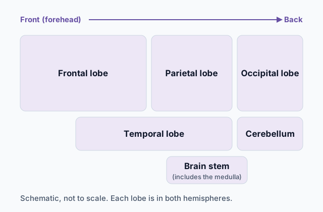 Tap to open the picture full size
Part 1 Think first A fall on the back of the head
Guess before you learn. A wrong guess still makes the answer stick.
Predict first
Lena, a cyclist, falls and hits the back of her head on the pavement. A doctor checks her eyes and finds nothing wrong with them, yet for weeks she has patches of her view where she cannot see clearly. Why would a blow to the back of the head affect her sight?
In short. The brain is built from structures that each generally lean toward certain jobs, and researchers work out those jobs with scans, case studies and surgical procedures.
- Structures low in the brain keep basic functions, alertness, coordination and balance running, while the lobes of the cerebral cortex handle sensing, language, movement and higher-order thinking.
- The two hemispheres are joined by the corpus callosum, and split-brain research shows they can specialize, with the language areas typically in the left hemisphere.
- Brain plasticity lets the brain rewire itself, so a different part can take over the function of a damaged one.
Part 2 Learn it Four short ideas, each checked as you go
2a. Keeping you alive, alert and steady
The structures low in the brain run jobs you rarely notice. The brain stem, which includes the medulla, generally controls basic functioning such as breathing and heart rate. You can hold your breath on purpose for a while, but once you stop trying, breathing carries on without you, and your heart keeps beating all night while you sleep.
Running up through the brain stem is the reticular activating system. Together with the brain's reward center, it generally controls alertness, some voluntary movement, eye movement, and some types of learning, cognition and emotion. When a sudden crash snaps a dozing student wide awake, alertness is the job in play. When something feels good and you find yourself wanting to do it again, the reward center is part of why that feeling teaches you anything.
At the back of the brain, below the rest, sits the cerebellum. It generally controls coordination of muscle movement, balance, and some forms of procedural learning: skills learned by doing, such as typing, until they run without step-by-step thought. Catching a ball and staying upright on an icy sidewalk both lean on it.
Notice the word generally in each of these. Brain structures share work, so a structure is linked to a job without being the only place that job happens.
Quick check · Sort it
Tap an example, then tap the structure that generally controls it.
Brain stem (medulla)
Reticular activating system and reward center
Cerebellum
2b. The cortex and its four lobes
The cerebral cortex is the brain's wrinkled outer layer, and it is divided into two halves, the left and right hemispheres. Along with it come the corpus callosum, a thick band of fibers joining the two hemispheres, and the limbic system, a group of structures made up of the thalamus, hypothalamus, pituitary gland, hippocampus and amygdala. Some textbooks draw the limbic system smaller and leave out the thalamus or the pituitary gland; the AP course lists all five, so count all five here. Later topics come back to several of these.
Each hemisphere has four lobes, and each lobe generally leans toward certain work.
- The occipital lobes, at the rear of the brain, handle visual information.
- The temporal lobes, on the sides, handle auditory and linguistic processing.
- The parietal lobes, near the back crown, hold the somatosensory cortex, which processes touch, and association areas, which process and organize information.
- The frontal lobes, just behind the forehead, handle linguistic processing, higher-order thinking and executive functioning, such as planning ahead and holding back an impulse. That work centers on the prefrontal cortex, at the very front. The motor cortex, at the rear of the frontal lobes, controls most types of skeletal movement.
To decide where to look, name the job first. Seeing points to the occipital lobes, hearing to the temporal lobes, touch to the parietal lobes, and moving or planning to the frontal lobes.
Quick check · Match
Tap a part of the cortex, then tap the activity it generally handles.
2c. Two hemispheres, and cutting the link
The corpus callosum lets the hemispheres share information. Severing it has been used to treat severe epilepsy, and research with those patients, called split-brain patients, shows that the right and left hemispheres may specialize in different activities and functions.
The best-known specialization is language, whose areas are typically in the left hemisphere. Broca's area is responsible for speech production, and Wernicke's area is responsible for speech comprehension. Damage to either can lead to aphasia, a loss of language ability. After damage to Broca's area, a person tends to understand speech but struggle to produce it. After damage to Wernicke's area, speech may flow but make little sense, and understanding others is hard.
To test specialization, researchers use the brain's contralateral organization: each hemisphere mostly handles the opposite side. The right hemisphere feels and moves the left side of the body, and the left hemisphere the right. In vision, everything to the left of the point you are looking at, your left visual field, goes to the right hemisphere, and your right visual field goes to the left hemisphere. That is true for both eyes, not one eye per side.
So a researcher has a split-brain patient stare at a dot and flashes a word beside it. A word flashed to the right of the dot reaches the left hemisphere, where the language areas usually are, and the patient reads it aloud. A word flashed to the left reaches only the right hemisphere.
Quick check · Put it in order
A split-brain patient stares at a dot while a picture of a key flashes briefly to the left of it. Tap the steps in the order they happen.
2d. A brain that rewires, and how we know
Brain plasticity is the brain's ability to rewire itself, modifying or creating new connections throughout development. It is also why a person can regain an ability after brain damage: the function of a damaged part can be assumed by a different part. The damaged tissue does not grow back as it was. Other areas take on the work, often slowly and with practice.
Researchers study how the brain works in several ways.
- An EEG records the brain's electrical activity through electrodes on the scalp. It shows when activity changes, moment by moment, but only roughly where.
- An fMRI scan tracks changes in blood flow to show which areas grow more active during a task.
- A case study looks in depth at one person, such as someone with damage to one area. It can show what is possible, but one person may not be typical.
- Lesioning is a surgical procedure that destroys a small area of brain tissue, usually in animals, so researchers can watch what changes. Because the researcher chooses the area, it can show what that area is needed for.
A scan showing an area active during a task fits the idea that the area does the work, but it does not show the task depends on it. Damage, whether from an injury or a lesion, shows what happens without the area.
Quick check · Match
Tap a research method, then tap the study that uses it.
Part 3 Watch it done Naming the damaged area, one question at a time
Watch each step, then do the last one yourself.
Grace, a woman who had a stroke, follows spoken instructions such as "point to the window" without trouble, and when a doctor tests her lips and tongue, they move normally. When she talks, though, she speaks in short, halting bursts with great effort: "Walk... dog... park." Damage to which area fits her changes best?
Step 1 · List what still works
She follows spoken instructions, so she hears and understands speech. Her lips and tongue move normally, so the muscles of speech and the commands that move them are working too.
Step 2 · Name what is lost
Getting words out takes great effort, and her sentences come out broken. What she has lost is producing speech, not understanding it. A loss of language ability after brain damage is aphasia, and language areas are typically in the left hemisphere.
Why this step?
Why separate producing speech from understanding it before naming an area?
Because the two language areas split that work. One is responsible for producing speech and the other for understanding it, so knowing which ability survived tells you which area the damage fits.
Your step · Decide and justify
Finish the reasoning. Grace, who had a stroke, understands spoken instructions, and her lips and tongue move normally when tested, but she speaks in short, halting bursts with great effort. Damage to which area fits best?
Which reason supports your answer?
Part 4 Try it Read a study: does another area take over?
Illustrative study (made up for practice, not a published study). Researchers recruited 48 adults. Twenty-four had regained much of their speech after a stroke damaged a language area in the left hemisphere, and 24 others of similar ages had never had a brain injury. People were placed in a group by their medical history; nobody can be assigned to have a stroke. Each adult named pictures aloud inside an fMRI scanner, and the researchers counted who showed strong activity in the matching area of the right hemisphere. A statistical test showed the difference between the groups was statistically significant.
| Group | Strong activity | Not strong | Total |
|---|---|---|---|
| Recovered after a stroke | 18 | 6 | 24 |
| No brain injury | 3 | 21 | 24 |
Try it · Calculate
In an illustrative fMRI study, 18 of 24 adults who recovered speech after a left-hemisphere stroke showed strong right-hemisphere activity while naming pictures, compared with 3 of 24 adults with no brain injury. By how many percentage points does the stroke group's percent exceed the other group's?
Try it · Decide and justify
In the illustrative study, 75% of adults who recovered speech after a left-hemisphere stroke showed strong right-hemisphere activity while naming pictures, against 12.5% of adults with no brain injury, a statistically significant difference. Groups were formed by medical history. A classmate says the study proves the right hemisphere took over their language work. Does it prove that?
Which reason supports your answer?
Part 5 Explain it Write it the way the exam asks
Nothing you type leaves this page.
Free response is 33.3% of the exam score. This one is shaped like Question 2, Evidence-Based Question.
Free-response practice · Question 2 style
Read the three sources, then answer the question they share: how does practicing a skill lost to brain damage relate to how much of that skill comes back? In parts B and C, cite the source you use, for example (Source 1).
Source 1. Illustrative study (made up for practice, not a published study). Researchers made a small lesion in the part of the motor cortex that controls one front paw in each of 40 rats, which left the rats clumsy at reaching for food with that paw. A random draw then put each rat in one of two groups of 20. One group practiced reaching for food pellets for 20 minutes a day; the other had no reaching practice. After six weeks, rats in the practice group grabbed the pellet on 68% of tries, compared with 41% for rats with no practice. The difference was statistically significant.
Source 2. Illustrative study (made up for practice, not a published study). Researchers followed 150 adults with aphasia after a stroke. For each adult they recorded the hours of speech therapy attended in the first six months and the gain on a speaking test over the same months. Adults who attended more hours of therapy tended to show larger gains, a positive correlation of r = +0.42.
Source 3. Illustrative case study (made up for practice, not a published study). A 34-year-old man had a stroke that damaged part of the motor cortex in his left hemisphere, leaving his right hand weak. He practiced hand exercises every day with a therapist. After eight months he could button a shirt with his right hand again. fMRI scans taken while he moved that hand showed activity in areas next to the damaged one that had not been active that way soon after the stroke.
(A) Propose a specific and defensible claim, based in psychological science, that answers the question.
(B) (i) Support your claim using one piece of specific and relevant evidence from one of the sources. (ii) Explain how the evidence from part B (i) supports your claim, using a psychological perspective, theory, concept, or research finding.
(C) (i) Support your claim using a piece of specific and relevant evidence from a different source. (ii) Explain how the evidence from part C (i) supports your claim, using a different psychological perspective, theory, concept, or research finding from the one in part B (ii).
Model answer
(A) The more a skill lost to brain damage is practiced, the more of that skill comes back.
(B) (i) In Source 1, rats that practiced reaching after a motor cortex lesion grabbed the pellet on 68% of tries, compared with 41% for rats that did not practice. (ii) This fits brain plasticity, the brain's ability to rewire itself so that a different part takes over the function of a damaged one. A random draw formed the groups, so practice is the likely reason the trained rats regained more reaching, which supports the claim that more practice brings back more of a lost skill.
(C) (i) In Source 2, adults with aphasia who attended more hours of speech therapy tended to gain more on a speaking test, a positive correlation of r = +0.42. (ii) This fits what aphasia is, a different concept from part B: a loss of language ability after damage to the brain's language areas, such as Broca's area, which is responsible for speech production. The speaking test measures exactly the ability these adults lost, so larger gains with more hours of therapy mean more of the lost skill returned with more practice, as the claim says, though a correlation alone cannot show the therapy caused the gains.
Score your own answer. This is a self-check, not a grade. Tick each point your answer makes.
Part 6 Check yourself Three people after a brain injury
On the exam, some multiple-choice questions share one set of information, the way these do.
Illustrative cases (made up for practice). Each row describes a fictional person, what happened to them, and what changed afterward.
| Person | What happened | What changed afterward |
|---|---|---|
| Dana | Had a stroke | Speaks easily in long sentences, but her words often make little sense, and she has trouble understanding what people say to her. Her hearing tests normal. |
| Mina | Had surgery to remove a small growth in her brain | With her eyes closed, she cannot tell where on her left hand she is being touched. She moves that hand normally. |
| Sam | Was injured in a car crash | Sees, speaks and moves normally, but acts on impulses and struggles to plan ahead. |
Case question 1 · Multiple choice
In a table of illustrative cases, Dana, a woman who had a stroke, speaks easily in long sentences, but her words often make little sense, and she has trouble understanding what people say to her. Her hearing tests normal. Damage to which area fits her changes best?
Case question 2 · Multiple choice
In a table of illustrative cases, Mina had surgery to remove a small growth in her brain. With her eyes closed, she cannot tell where on her left hand she is being touched, though she moves that hand normally. Damage in which area fits her changes best?
Case question 3 · Decide and justify
In a table of illustrative cases, Sam was injured in a car crash. He sees, speaks and moves normally, but acts on impulses and struggles to plan ahead. A scan shows the injury is in his prefrontal cortex. A classmate says Sam's case proves the prefrontal cortex controls planning in every person. Is the classmate right?
Which reason supports your answer?
The rest stand on their own and do not use the case.
Check yourself · Sort it
Tap each structure, then tap where it belongs.
Part of the limbic system
Not part of the limbic system
Check yourself · Spot the error
In an illustrative case, Rosa, a woman who had a stroke in her left hemisphere, lost much of her speech and regained a good part of it over a year of daily therapy. A student explains her recovery. Tap the first line that goes wrong.
Check yourself · Fill the statement
Tap a blank, then tap a word.
To treat severe epilepsy, a surgeon may sever the , the band of fibers that joins the two hemispheres. Near the back crown, the parietal lobes hold the cortex, which processes touch, and areas, which process and organize information. Planning ahead and holding back an impulse are executive work of the lobes. Damage to Broca's or Wernicke's area can lead to , a loss of language ability.
Part 7 Keep it Bring back earlier ideas
Coming back to older ideas is how they stay usable on the exam.
Bring it back · Multiple choice
Jae presses a fingertip lightly, then much harder, on a pressure sensor. Whenever the stimulation reaches a sensory neuron's threshold, the neuron fires, and each action potential is the same size; pressing harder never makes a single action potential bigger. Which idea does this show?
Bring it back · Match
Tap a kind of study, then tap the design that fits it.
In three lines
- The brain stem, reticular activating system, reward center and cerebellum handle basic functioning, alertness and coordination, and each lobe of the cortex leans toward its own kind of work.
- Split-brain research uses each visual field's path to the opposite hemisphere to show the hemispheres can specialize, with Broca's and Wernicke's areas typically on the left.
- Brain plasticity lets a different part take over for a damaged one, and EEG, fMRI, case studies and lesioning each show something different about how the brain works.
Vocabulary
- Brain stem
- The structure, including the medulla, that generally controls basic functioning such as breathing and heart rate.
- Reticular activating system
- A system that, with the reward center, generally controls alertness, some voluntary movement, eye movement, and some types of learning, cognition and emotion.
- Reward center
- Part of the brain that, with the reticular activating system, is linked to some types of learning, cognition and emotion.
- Cerebellum
- The structure that generally controls coordination of muscle movement, balance, and some forms of procedural learning.
- Cerebral cortex
- The brain's wrinkled outer layer, divided into two hemispheres and made up of the lobes. The corpus callosum and the limbic system are grouped with it.
- Limbic system
- The group of structures made up of the thalamus, hypothalamus, pituitary gland, hippocampus and amygdala.
- Corpus callosum
- The band of fibers that joins the brain's two hemispheres.
- Occipital lobes
- Lobes at the rear of the brain that generally control visual information processing.
- Temporal lobes
- Lobes on the sides of the brain that generally control auditory and linguistic processing.
- Parietal lobes
- Lobes near the back crown of the brain that generally control association areas and the somatosensory cortex.
- Association areas
- Areas of the parietal lobes that process and organize information.
- Somatosensory cortex
- The area of the parietal lobes that processes touch sensitivity.
- Frontal lobes
- Lobes just behind the forehead that generally control linguistic processing, higher-order thinking and executive functioning.
- Prefrontal cortex
- The front part of the frontal lobes, where higher-order thinking and executive functioning are especially centered.
- Motor cortex
- The area at the rear of the frontal lobes that controls most types of skeletal movement.
- Split-brain research
- Research with people whose corpus callosum has been severed, often to treat severe epilepsy, showing that the two hemispheres may specialize.
- Contralateral hemispheric organization
- The arrangement in which each hemisphere mostly handles the opposite side, so each visual field goes to the opposite hemisphere.
- Broca's area
- An area, typically in the left hemisphere, responsible for speech production.
- Wernicke's area
- An area, typically in the left hemisphere, responsible for speech comprehension.
- Aphasia
- A loss of language ability that can follow damage to the brain's language areas.
- Brain plasticity
- The ability of the brain to rewire itself or modify or create new connections throughout development, which generally lets a different part take over the function of a damaged part.
- EEG
- A scan that records the brain's electrical activity through electrodes on the scalp.
- fMRI
- A scan that tracks changes in blood flow to show which brain areas are more active during a task.
- Case study
- An in-depth study of one person, such as someone with damage to one brain area.
- Lesioning
- A surgical procedure that destroys a small area of brain tissue, usually in animals, to see what changes.
Course alignment, for teachers
AP Psychology topic 1.4, Unit 1: Biological Bases of Behavior.
- Skill 1.A: Apply psychological perspectives, theories, concepts, and research findings to a scenario.
- Skill 2.C: Evaluate the appropriate use of research design elements in non-experimental methodologies.
- Skill 2.D: Evaluate whether a psychological research scenario followed appropriate ethical procedures.
- Skill 3.A: Identify psychology-related concepts in descriptions or representations of data.
This lesson maps all 13 essential knowledge statements of the topic to the part that teaches each one: 13 are checked by a scored question. The map is in the lesson file.